FOLLOW US

This is default featured slide 1 title

Go to Blogger edit html and find these sentences.Now replace these sentences with your own descriptions.This theme is Bloggerized by Lasantha Bandara - Premiumbloggertemplates.com.

This is default featured slide 2 title

Go to Blogger edit html and find these sentences.Now replace these sentences with your own descriptions.This theme is Bloggerized by Lasantha Bandara - Premiumbloggertemplates.com.

This is default featured slide 3 title

Go to Blogger edit html and find these sentences.Now replace these sentences with your own descriptions.This theme is Bloggerized by Lasantha Bandara - Premiumbloggertemplates.com.

This is default featured slide 4 title

Go to Blogger edit html and find these sentences.Now replace these sentences with your own descriptions.This theme is Bloggerized by Lasantha Bandara - Premiumbloggertemplates.com.

This is default featured slide 5 title

Go to Blogger edit html and find these sentences.Now replace these sentences with your own descriptions.This theme is Bloggerized by Lasantha Bandara - Premiumbloggertemplates.com.

Showing posts with label surgery. Show all posts

Saturday, February 6, 2016

Amebic liver abscess

Amebic liver abscess :
is the most frequent extraintestinal manifestation ofEntamoeba histolytica infection. This infection is caused by the protozoa E histolytica, which enter the portal venous system from the colon. Amebic liver abscess is an important cause of space-occupying lesions of the liver, mainly in developing countries. Prompt recognition and appropriate treatment of amebic liver abscess lead to improved morbidity and mortality.

Pathophysiology

E histolytica exists in 2 forms. The cyst stage is the infective form, and the trophozoite stage causes invasive disease. People who chronically carry E histolytica shed cysts in their feces; these cysts are transmitted primarily by food and water contamination. Rare cases of transmission via oral and anal sex or direct colonic inoculation through colonic irrigation devices have occurred. Cysts are resistant to gastric acid, but the wall is broken down by trypsin in the small intestine. Trophozoites are released and colonize the cecum. To initiate symptomatic infection, E histolytica trophozoites present in the lumen must adhere to the underlying mucosa and penetrate the mucosal layer.
Liver involvement occurs following invasion of E histolytica into mesenteric venules. Amebae then enter the portal circulation and travel to the liver where they typically form large abscesses. The Gal/GalNAc lectin is an adhesion protein complex that sustains tissue invasion.[1] The abscess contains acellular proteinaceous debris, which is thought to be a consequence of induced apoptosis[2] and is surrounded by a rim of amebic trophozoites invading the tissue.
The right lobe of the liver is more commonly affected than the left lobe. This has been attributed to the fact that the right lobe portal laminar blood flow is supplied predominantly by the superior mesenteric vein, whereas the left lobe portal blood flow is supplied by the splenic vein.

Epidemiology

Frequency

United States
Amebic liver abscess is rare and is currently seen almost exclusively in immigrants or travelers. In 1994, 2,983 cases of amebiasis were reported to the Centers for Disease Control (CDC). The disease was removed from the National Notifiable Diseases Surveillance System in 1995. An estimated 4% of patients with amebic colitis develop an amebic liver abscess.
An estimated 10% of the population is infected with Entamoeba dispar. Previously thought to be a nonpathogenic strain of E histolytica, this type of amoeba does not produce clinical symptoms even in the immunocompromised host.
Race-, sex-, and age-related demographics
All races can be affected by amebic liver abscess. Risk factors for infection include travel or residence in endemic areas.
Amebic liver abscess is marked by a 7-12–fold higher incidence in males than in females despite an equal sex distribution of noninvasive colonic amebic disease among adults.[3] However, no sexual preponderance exists among children.
Peak incidence of amebic liver abscess occurs in people in their third, fourth, and fifth decades of life, although it can occur in any age group.
International
Worldwide, approximately 40-50 million people are infected annually, with the majority of infections occurring in developing countries. The prevalence of infection is higher than 5-10% in endemic areas[4] and sometimes as high as 55%.[5] The highest prevalence is found in developing countries in the tropics, particularly in Mexico, India, Central and South America, and tropical areas of Asia and Africa.

Mortality/Morbidity

Infection with E histolytica ranks second worldwide among parasitic causes of death, following malaria.
Annually, 40,000-100,000 deaths are caused by infection with E histolytica. Per year, a 10% risk of developing symptomatic invasive amebiasis exists after the acquisition of a pathogenic strain.[6]
Complications
Pleuropulmonary infection is the most common complication. Mechanisms of infection include development of a sympathetic serous effusion; rupture of a liver abscess into the chest cavity, leading to empyema; or hematogenous spread, resulting in parenchymal infection.
Bronchopleural fistula may occur in rare instances when patients expectorate a substance that resembles anchovy paste. Trophozoites may be demonstrated in the fluid. Occasionally, this complication may be followed by a spontaneous cure of the amebic liver abscess.
Cardiac involvement results following the rupture of an abscess involving the left lobe of the liver. It usually is associated with very high mortality.
Intraperitoneal rupture occurs in 2-7% of patients. Left lobe abscesses are more likely to progress to rupture because of their later clinical presentation.
Bacterial superinfection can occur.
Rupture into peritoneal organs (eg, stomach) and mediastinum can occur.
Cases of hepatic artery pseudoaneurysm have been reported.

History

The signs and symptoms of amebic liver abscess often are nonspecific, resembling those of pyogenic liver abscess or other febrile diseases.[7, 8, 9, 10]

Time of onset

Patients with amebic liver abscess usually present acutely (duration of symptoms < 14 d), with the most frequent complaints being fever and abdominal pain. This presentation is characteristic of younger patients.
The subacute presentation is characterized by weight loss, and, in less than half the cases, abdominal pain and fever are present.

Abdominal pain

Abdominal pain is the most common element in the history and is present in 90-93% of patients. The pain is usually constant, dull, and aching, and it is most frequently located in the right upper quadrant (54-67%) and may radiate to the right shoulder or scapular area.
The pain increases with coughing, walking, and deep breathing, as well as when patients rest on their right side.

Constitutional symptoms

Fever is present in 87-100% of cases, and rigors are present in 36-69% of cases.
Nausea and vomiting are present in 32-85% of cases, and weight loss is present in 33-64% of cases.

Diarrhea

Diarrhea is present in less than one third of patients at the time of diagnosis. Some patients describe a history of having had dysentery within the previous few months. Bloody diarrhea is present in 7% of cases.

Pulmonary symptoms

Pulmonary symptoms are present in 18-26% of cases. The most frequent symptoms are cough and chest pain, which may represent a sign of secondary pulmonary involvement by abscess rupture in the pleural cavity.
When coughing produces an odorless brown substance similar to anchovy paste, a bronchopleural fistula has developed.[11]

Recent travel to endemic areas

Onset of symptoms usually occurs within 8-12 weeks from the date of travel. In 95% of cases, onset occurs within 5 months of returning from travel to an endemic area. A remote travel history of as many as 12 years has been reported.

Physical

Fever is the most common sign and is found in as many as 99% of cases.
Hepatomegaly is present in some cases. The frequency varies widely in different series published, reporting as high as 63% in one series and as low as 18% in another.
Hepatomegaly with pain upon palpation is one of the most important signs of amebic liver abscess. Point tenderness over the liver, below the ribs, or in the intercostal spaces is a typical finding.
Right upper abdominal quadrant tenderness is present in 55-75% of cases. When the abscess is located in the left lobe (28% of cases), epigastric tenderness is noted.
Pulmonary abnormalities are present in 20-45% of cases, and they consist of dullness and rales at the right lung base and nonproductive cough. Breath sounds over the right lung base may be diminished. Pleural rub may be audible.
Jaundice (< 10% of cases) most often occurs in complicated cases with multiple abscesses or a large abscess compressing the biliary tract.
Signs of complications include the following:
  • Signs of peritoneal irritation, such as rebound tenderness, guarding, and absence of bowel sounds, are present when the abscess ruptures into the peritoneal cavity. Peritonitis occurs in 2-7% of cases.
  • Pericardial friction rub can be audible when the abscess extends into the pericardium. This sign is associated with a very high mortality.
  • Signs of pleural effusion are present when the abscess ruptures into the pleural cavity.

Causes

The following are the risk factors associated with amebic liver abscess:
  • Immigrants from endemic areas
  • Institutionalized persons, especially people with mental retardation
  • Crowding and poor hygiene
  • Men who have sex with men (secondary to sexually acquired amebic colitis)
  • Presence of immunosuppression (eg, HIV infection, malnutrition with hypoalbuminemia, alcohol abuse, chronic infections, posttraumatic splenectomy, steroid use) 
  • Medical Care

    Most uncomplicated amebic liver abscesses can be treated successfully with amebicidal drug therapy alone. Use tissue amebicides to eradicate the invasive trophozoite forms in the liver. After completion of treatment with tissue amebicides, administer luminal amebicides for eradication of the asymptomatic colonization state. Failure to use luminal agents can lead to relapse of infection in approximately 10% of patients.
    In general, metronidazole, tinidazole, emetine, and dehydroemetine are active in invaded tissues; chloroquine is active only in the liver; tetracycline acts on the bowel wall; and diloxanide furoate, paromomycin, and iodoquinol are luminal agents only. The details on tissue and luminal amebicidal agents are discussed in Medication.

    Metronidazole

    Metronidazole remains the drug of choice for amebic liver abscess. Metronidazole enters the protozoa by passive diffusion and is converted to reactive cytotoxic nitroradicals by reduced ferredoxin or flavodoxin. Tinidazole, another nitroimidazole closely related to metronidazole, was approved for the treatment of amebic liver abscess and invasive amebiasis. Tinidazole is well tolerated by patients. Tinidazole may be administered once daily and appears to be at least as effective as metronidazole, with a clinical cure rate of more than 90%.
    Metronidazole, 750 mg 3 times a day orally for 10 days, was reported to be curative in 90% of patients with amebic liver abscess. The drug also is available for intravenous administration for those patients who are unable to take medication by the oral route.
    Resolution of symptoms is fairly rapid and is observed within 3 days in most patients in the United States. In endemic areas outside the United States, it takes relatively longer to resolve symptoms because the abscesses are quite large or multiple by the time patients seek medical attention.
    In vivo resistance to metronidazole by E histolytica has not been reported. Nevertheless, in vitro studies have shown an association between metronidazole resistance and decreased expression of ferredoxin 1 and flavodoxin and increased expression of iron-containing superoxide dismutase and peroxiredoxin in E histolytica.
    Usual adverse effects of metronidazole include nausea, headache, and metallic taste. Abdominal cramps, vomiting, diarrhea, and dizziness also may occur. Dark urine may occur from a metabolite of the drug.

    Other items to consider

    Other considerations include the following:
    • No randomized controlled trials exist that demonstrate the benefits of combination therapy over monotherapy.
    • Outside the United States, other closely related amebicidal agents, such as secnidazole or ornidazole, can be substituted in appropriate dosages. These drugs are not available in the United States.
    • Chloroquine phosphate may be substituted or added in the event of failure of resolution of clinical symptoms with metronidazole or another nitroimidazole within 5 days or intolerance to metronidazole or a nitroimidazole. Chloroquine has the disadvantage of being associated with higher relapse rates than nitroimidazoles. Adverse effects include gastrointestinal upset, headache, dizziness, and blurred vision. Retinopathy does not occur at the dose used for amebic liver abscess.
    • Emetine or dehydroemetine has a direct lethal action on the trophozoites ofE histolytica. These agents are very toxic and, therefore, should be used only as a second-line therapy. Their toxicity includes cardiac arrhythmias, precordial pain, muscle weakness, vomiting, and diarrhea. Dehydroemetine is less toxic than emetine.
    Administer a luminal amebicidal agent to eradicate the intestinal carriage after the amebic liver abscess has been treated with one of the above tissue amebicides. Failure to use luminal agents can lead to relapse of infection in approximately 10% of patients. Luminal agents with proven efficacy include diloxanide furoate, iodoquinol, and paromomycin. Note the following:
    • Diloxanide furoate is free of major adverse effects. The most common adverse effect is flatulence and occasional gastrointestinal upset.
    • Iodoquinol (diiodohydroxyquin) rarely causes abdominal pain, diarrhea, or rash. A structurally related diiodohydroxyquin caused subacute myelopticoneuropathy and is obsolete now.
    • Although paromomycin may occasionally cause nausea, abdominal cramps, or diarrhea, it is the preferred luminal amebicidal.
    • The details on the luminal amebicides are discussed in Medication.

    Consultations

    Consult with an interventional radiologist for imaging-guided aspiration of the abscess.
    Consult with a general surgeon for open surgical drainage of the abscess under rare circumstances (see Surgical Care).

    Diet and activity

    No specific diet change or modification is required. However, discuss food hygiene with patients because amebiasis is associated with suboptimal personal or food hygiene (see Deterrence/Prevention).
    No restriction of activity is needed, except during the first few days of acute illness with pain.
    If emetine or dehydroemetine is used, the patient should remain sedentary for approximately 4 weeks after completing therapy because of their toxicity.

    Surgical Care

    Consider therapeutic aspiration of amebic liver abscess in the following situations: (1) high risk of abscess rupture, as defined by cavity size greater than 5 cm; (2) left lobe liver abscess, which is associated with higher mortality and frequency of peritoneal leak or rupture into the pericardium; (3) failure to observe a clinical medical response to therapy within 5-7 days; and (4) cannot differentiate from a pyogenic liver abscess.
    The following are predictive of the need for aspiration: (1) age older than 55 years, (2) abscess greater than 5 cm in diameter, and (3) failure of medical therapy after 7 days.[25] In endemic areas, because of the late presentation and the existence of multiple abscesses, as many as 50% of patients may require aspiration.[26]However, routine needle aspiration offers only minimal benefit over medical care alone for uncomplicated amebic liver abscess and, unless one of the above indications exists, should be avoided.[27] Prompt medical care decreases the need for aspiration.[28]
    Imaging-guided needle aspiration and catheter drainage are the procedures of choice. Generally, surgical drainage is not necessary and should be avoided; however, consider open surgical drainage when the abscess is inaccessible to needle drainage or a response to therapy has not occurred in 5-7 days.
    Simple needle aspiration is less invasive, is less expensive, and has the advantage of being able to drain multiple abscesses in the same session. Simple needle aspiration avoids problems related to catheter care (see Procedures).
    Although catheter drainage may be more effective than needle aspiration, in a study by Rajak et al,[29] the average time for clinical improvement, mean hospital stay, and time to resolution were similar among the patients who were successfully treated in the 2 treatment groups.

Saturday, January 23, 2016

Liver abscess

Background

Bacterial abscess of the liver is relatively rare; however, it has been described since the time of Hippocrates (400 BC), with the first published review by Bright appearing in 1936. In 1938, Ochsner's classic review heralded surgical drainage as the definitive therapy; however, despite the more aggressive approach to treatment, the mortality remained at 60-80%.[1]
The development of new radiologic techniques, the improvement in microbiologic identification, and the advancement of drainage techniques, as well as improved supportive care, have reduced mortality to 5-30%; yet, the prevalence of liver abscess has remained relatively unchanged. Untreated, this infection remains uniformly fatal.
The three major forms of liver abscess, classified by etiology, are as follows:
  • Pyogenic abscess, which is most often polymicrobial, accounts for 80% of hepatic abscess cases in the United States
  • Amebic abscess due to Entamoeba histolytica accounts for 10% of cases [2]
  • Fungal abscess, most often due to Candida species, accounts for fewer than 10% of cases
For patient education resources, see the Infections Center and the Digestive Disorders Center, as well as Skin Abscess and Antibiotics.

Pathophysiology

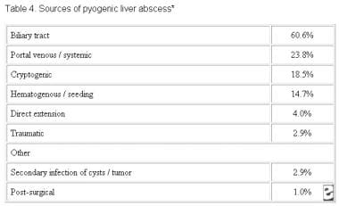
The liver receives blood from both systemic and portal circulations. Increased susceptibility to infections would be expected given the increased exposure to bacteria. However, Kupffer cells lining the hepatic sinusoids clear bacteria so efficiently that infection rarely occurs. Multiple processes have been associated with the development of hepatic abscesses (see the image below).
Table 4: Underlying etiology of 1086 cases of liveTable 4: Underlying etiology of 1086 cases of liver abscess compiled from the literature.
Appendicitis was traditionally the major cause of liver abscess. As diagnosis and treatment of this condition has advanced, its frequency as a cause for liver abscess has decreased to 10%.
Biliary tract disease is now the most common source of pyogenic liver abscess(PLA). Obstruction of bile flow allows for bacterial proliferation. Biliary stone disease, obstructive malignancy affecting the biliary tree, stricture, and congenital diseases are common inciting conditions. With a biliary source, abscesses usually are multiple, unless they are associated with surgical interventions or indwelling biliary stents. In these instances, solitary lesions can be seen.
Infections in organs in the portal bed can result in a localized septic thrombophlebitis, which can lead to liver abscess. Septic emboli are released into the portal circulation, trapped by the hepatic sinusoids, and become the nidus for microabscess formation. These microabscesses initially are multiple but usually coalesce into a solitary lesion.
Microabscess formation can also be due to hematogenous dissemination of organisms in association with systemic bacteremia, such as endocarditis and pyelonephritis. Cases also are reported in children with underlying defects in immunity, such as chronic granulomatous disease and leukemia.
Approximately 4% of liver abscesses result from fistula formation between local intra-abdominal infections.
Despite advances in diagnostic imaging, cryptogenic causes account for a significant proportion of cases; surgical exploration has impacted this minimally. These lesions usually are solitary in nature.
Penetrating hepatic trauma can inoculate organisms directly into the liver parenchyma, resulting in pyogenic liver abscess. Nonpenetrating trauma can also be the precursor to pyogenic liver abscess by causing localized hepatic necrosis, intrahepatic hemorrhage, and bile leakage. The resulting tissue environment permits bacterial growth, which may lead to pyogenic liver abscess. These lesions are typically solitary.
PLA has been reported as a secondary infection of amebic abscess, hydatid cystic cavities, and metastatic and primary hepatic tumors. It is also a known complication of liver transplantation, hepatic artery embolization in the treatment of hepatocellular carcinoma, and the ingestion of foreign bodies, which penetrate the liver parenchyma. Trauma and secondarily infected liver pathology account for a small percentage of liver abscess cases.
The right hepatic lobe is affected more often than the left hepatic lobe by a factor of 2:1. Bilateral involvement is seen in 5% of cases. The predilection for the right hepatic lobe can be attributed to anatomic considerations. The right hepatic lobe receives blood from both the superior mesenteric and portal veins, whereas the left hepatic lobe receives inferior mesenteric and splenic drainage. It also contains a denser network of biliary canaliculi and, overall, accounts for more hepatic mass. Studies have suggested that a streaming effect in the portal circulation is causative.

Etiology

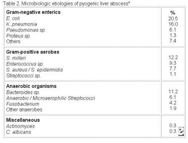
Polymicrobial involvement is common, with Escherichia coli and Klebsiella pneumoniae being the two most frequently isolated pathogens (see the image below). Reports suggest that K pneumoniae is an increasingly prominent cause.[4]
Table 2: Microbiologic results from 312 cases of lTable 2: Microbiologic results from 312 cases of liver abscess compiled from the literature.
Enterobacteriaceae are especially prominent when the infection is of biliary origin. Abscesses involving K pneumoniae have been associated with multiple cases of endophthalmitis.
The pathogenic role of anaerobes was underappreciated until the isolation of anaerobes from 45% of cases of pyogenic liver abscess was reported in 1974. Since that time, increasing rates of anaerobic involvement have been reported, likely because of increased awareness and improved culturing techniques. The most frequently encountered anaerobes are Bacteroides species, Fusobacteriumspecies, and microaerophilic and anaerobic streptococci. A colonic source is usually the initial source of infection.
Staphylococcus aureus abscesses usually result from hematogenous spread of organisms involved with distant infections, such as endocarditis. S milleri is neither anaerobic nor microaerophilic. It has been associated with both monomicrobial and polymicrobial abscesses in patients with Crohn disease, as well as with other patients with pyogenic liver abscess.
Amebic liver abscess is most often due to E histolytica. Liver abscess is the most common extraintestinal manifestation of this infection.
Fungal abscesses primarily are due to Candida albicans and occur in individuals with prolonged exposure to antimicrobials, hematologic malignancies, solid-organ transplants, and congenital and acquired immunodeficiency. Cases involvingAspergillus species have been reported.
Other organisms reported in the literature include Actinomyces species, Eikenella corrodens, Yersinia enterocolitica, Salmonella typhi, and Brucella melitensis.
A small case series in Taiwan investigated pyogenic liver abscess as the initial manifestation of underlying hepatocellular carcinoma. In regions with a high prevalence of both pyogenic liver abscess and hepatocellular carcinoma, clinicians should be aware of the possibility of underlying hepatocellular carcinoma in patients with risk factors for the disease.[5]

Epidemiology

United States statistics

The incidence of pyogenic liver abscess has essentially remained unchanged by both hospital and autopsy data. Liver abscess was diagnosed in 0.7%, 0.45%, and 0.57% of autopsies during the periods of 1896-1933, 1934-1958, and 1959-1968, respectively. The frequency in hospitalized patients is in the range of 8-16 cases per 100,000 persons. Studies suggest a small, but significant, increase in the frequency of liver abscess.

Age-related demographics

Prior to the antibiotic era, liver abscess was most common in the fourth and fifth decades of life, primarily due to complications of appendicitis. With the development of better diagnostic techniques, early antibiotic administration, and the improved survival of the general population, the demographic has shifted toward the sixth and seventh decades of life. Frequency curves display a small peak in the neonatal period followed by a gradual rise beginning at the sixth decade of life.
Cases of liver abscesses in infants have been associated with umbilical vein catheterization and sepsis.
When abscesses are seen in children and adolescents, underlying immune deficiency, severe malnutrition, or trauma frequently exists.

Sex-related demographics

While abscesses once showed a predilection for males in earlier decades, no sexual predilection currently exists. Males have a poorer prognosis from hepatic abscess than females.

Prognosis

Untreated, pyogenic liver abscess remains uniformly fatal. With timely administration of antibiotics and drainage procedures, mortality currently occurs in 5-30% of cases. The most common causes of death include sepsis, multiorgan failure, and hepatic failure.[3]
Indicators of a poor prognosis have been described since 1938 and include multiplicity of abscesses, underlying malignancy, severity of underlying medical conditions, presence of complications, and delay in diagnosis.[3]
Indicators of a poor prognosis in amebic abscess include a bilirubin level of greater than 3.5 mg/dL, encephalopathy, hypoalbuminemia (ie, serum albumin level of <2 g/dL), and multiple abscesses; all are independent factors that predict poor outcome.
An underlying malignant etiology and an Acute Physiology and Chronic Health Evaluation (APACHE II) score greater than 9 increases the relative mortality by 6.3-fold and 6.8-fold, respectively.
Chen et al examined prognostic factors for elderly patients with pyogenic liver abscess.[9] Results from the study, which included 118 patients aged 65 years or older and 221 patients below age 65 years, indicated that age and an APACHE II score of 15 or greater at hospital admission were risk factors for mortality. The evidence ultimately suggested that outcomes for older patients with pyogenic liver abscess are on a par with those for younger patients. The investigators also found that in the younger patient group, there was greater frequency of males suffering from alcoholism, a cryptogenic abscess, and K pneumoniae infection.

History

The most frequent symptoms of hepatic abscess include the following (see the image below):
  • Fever (either continuous or spiking)
  • Chills
  • Right upper quadrant pain
  • Anorexia
  • Malaise
Table 1: Presenting symptoms and signs in 715 patiTable 1: Presenting symptoms and signs in 715 patients diagnosed with liver abscess.
Cough or hiccoughs due to diaphragmatic irritation may be reported. Referred pain to the right shoulder may be present.
Individuals with solitary lesions usually have a more insidious course with weight loss and anemia of chronic disease. With such symptoms, malignancy often is the initial consideration.
Fever of unknown origin (FUO) frequently can be an initial diagnosis in indolent cases. Multiple abscesses usually result in more acute presentations, with symptoms and signs of systemic toxicity.
Afebrile presentations have been documented.

Physical Examination

Fever and tender hepatomegaly are the most common signs. A palpable mass need not be present. Midepigastric tenderness, with or without a palpable mass, is suggestive of left hepatic lobe involvement.
Decreased breath sounds in the right basilar lung zones, with signs of atelectasis and effusion on examination or radiologically, may be present. A pleural or hepatic friction rub can be associated with diaphragmatic irritation or inflammation of Glisson capsule.
Jaundice may be present in as many as 25% of cases and usually is associated with biliary tract disease or the presence of multiple abscesses.

Complications

Complications of liver abscess may include the following:
  • Sepsis
  • Empyema resulting from contiguous spread or intrapleural rupture of abscess
  • Rupture of abscess with resulting peritonitis
  • Endophthalmitis when an abscess is associated with K pneumoniaebacteremia
  • Medical Care

    An untreated hepatic abscess is nearly uniformly fatal as a result of complications that include sepsis, empyema, or peritonitis from rupture into the pleural or peritoneal spaces, and retroperitoneal extension. Treatment should include drainage, either percutaneous or surgical.
    Antibiotic therapy as a sole treatment modality is not routinely advocated, though it has been successful in a few reported cases. It may be the only alternative in patients too ill to undergo invasive procedures or in those with multiple abscesses not amenable to percutaneous or surgical drainage. In these instances, patients are likely to require many months of antimicrobial therapy with serial imaging and close monitoring for associated complications.
    Antimicrobial treatment is a common adjunct to percutaneous or surgical drainage.

    Surgical Care

    Surgical drainage was the standard of care until the introduction of percutaneous drainage techniques in the mid-1970s. With the refinement of image-guided techniques, percutaneous drainage and aspiration have become the standard of care.
    Current indications for the surgical treatment of pyogenic liver abscess are for the treatment of underlying intra-abdominal processes, including signs of peritonitis; existence of a known abdominal surgical pathology (eg, diverticular abscess); failure of previous drainage attempts; and the presence of a complicated, multiloculated, thick-walled abscess with viscous pus.
    Shock with multisystem organ failure is a contraindication for surgery.
    Open surgery can be performed by either of the following two approaches:
    • A transperitoneal approach allows for abscess drainage and abdominal exploration to identify previously undetected abscesses and the location of an etiologic source
    • For high posterior lesions, a posterior transpleural approach can be used; although this affords easier access to the abscess, the identification of multiple lesions or a concurrent intra-abdominal pathology is lost
    A laparoscopic approach is also commonly used in select cases. This minimally invasive approach affords the opportunity to explore the entire abdomen and to significantly reduce patient morbidity. A growing literature is defining the optimal population for this mode of intervention.
    A retrospective chart review compared surgery versus percutaneous drainage for liver abscesses greater than 5 cm. Morbidity was comparable for the two procedures, but those treated with surgery had fewer secondary procedures and fewer treatment failures.
    Postoperative complications are not uncommon and include recurrent pyogenic liver abscess, intra-abdominal abscess, hepatic or renal failure, and wound infection.

    Consultations

    Obtain an interventional radiology consultation as soon as the diagnosis is considered to allow rapid collection of cavity fluid and the potential for early therapeutic drainage of abscess.
    Immediately seek a consultation with a general surgeon if the source of the abscess is a known underlying abdominal pathology or in cases with peritonitis. In cases undergoing percutaneous drainage, seek the involvement of a general surgeon if drainage of the abscess cavity is unsuccessful.
    Gastroenterology involvement may be useful after successful drainage to evaluate for underlying gastrointestinal disease using colonoscopy or endoscopic retrograde cholangiopancreatography (ERCP).
    Infectious disease consultation should be considered in complicated cases and when the involved pathogens are unusual or difficult to treat, such as in fungal abscesses.

    Long-Term Monitoring

    Aggressively seek an underlying source of the abdominal pathology.
    Perform weekly serial computed tomography (CT) or ultrasound examinations to document adequate drainage of the abscess cavity. Continue radiologic evaluation to document progress of therapy after discharge.
    Drain care may be required. Maintain drains until the output is less than 10 mL/day.
    Monitor fever curves. Persistent fever after 2 weeks of therapy may indicate the need for more aggressive drainage.
    For patients with an underlying malignancy, definitive treatment, such as surgical removal of the mass, should be pursued if at all possible.
    Patients will require prolonged parenteral antimicrobial therapy that may continue after discharge. Monitoring of medication levels, renal function, and blood counts may be needed. Enteral nutrition is the preferred route unless it is clinically contraindicated.
  • Medication Summary

    Until cultures are available, the choice of antimicrobial agents should be directed toward the most commonly involved pathogens. Regimens using beta-lactam/beta-lactamase inhibitor combinations, carbapenems, or second-generation cephalosporins with anaerobic coverage are excellent empiric choices for the coverage of enteric bacilli and anaerobes. Metronidazole or clindamycin should be added for the coverage of Bacteroides fragilis if other employed antibiotics offer no anaerobic coverage.
    Amebic abscess should be treated with metronidazole, which will be curative in 90% of cases. Metronidazole should be initiated before serologic test results are available. Patients who do not respond to metronidazole should receive chloroquine alone or in combination with emetine or dehydroemetine.
    Systemic antifungal agents should be initiated if fungal abscess is suspected and after the abscess has been drained percutaneously or surgically. Initial therapy for fungal abscess is currently amphotericin B. Lipid formulations may offer some benefit in that the complexing of drug to lipid moieties allows for concentration in hepatocytes. Further investigation is required for definitive proof. Cases of successful fluconazole treatment after amphotericin failure have been reported; however, its use as an initial agent is still being studied.
    Ultimately, the organisms isolated and antibiotic sensitivities should guide the final choice of antimicrobials.
    Duration of treatment has always been debated. Short courses (2 wk) of therapy after percutaneous drainage have been successful in a small series of patients; however, most series have reported recurrence of abscess even after more prolonged courses. Currently 4-6 weeks of therapy is recommended for solitary lesions that have been adequately drained. Multiple abscesses are more problematic and can require up to 12 weeks of therapy. Both the clinical and radiographic progress of the patient should guide the length of therapy.

    Antibiotics

    Class Summary

    Empiric antimicrobial therapy must be comprehensive and should cover all likely pathogens in the context of the clinical setting.

    Meropenem (Merrem)

    Bactericidal broad-spectrum carbapenem antibiotic that inhibits cell-wall synthesis. Effective against most gram-positive and gram-negative bacteria.
    Has slightly increased activity against gram negatives and slightly decreased activity against staphylococci and streptococci species compared to imipenem.

    Imipenem and cilastatin (Primaxin)

    For treatment of multiple organism infections in which other agents do not have wide-spectrum coverage or are contraindicated due to potential for toxicity.

    Cefuroxime (Ceftin)

    Second-generation cephalosporin maintains gram-positive activity that first-generation cephalosporins have; adds activity against Proteus mirabilis,Haemophilus influenzae, Escherichia coli, Klebsiella pneumoniae, and Moraxella catarrhalis. Condition of patient, severity of infection, and susceptibility of microorganism determine proper dose and route of administration.

    Cefotetan (Cefotan)

    Second-generation cephalosporin indicated for infections caused by susceptible gram-positive cocci and gram-negative rods.
    Dosage and route of administration depends on condition of patient, severity of infection, and susceptibility of causative organism.

    Cefoxitin (Mefoxin)

    Second-generation cephalosporin indicated for gram-positive cocci and gram-negative rod infections. Infections caused by cephalosporin-resistant or penicillin-resistant gram-negative bacteria may respond to cefoxitin.

    Cefaclor (Ceclor)

    Second-generation cephalosporin indicated for infections caused by susceptible gram-positive cocci and gram-negative rods.
    Determine proper dosage and route based on condition of patient, severity of infection, and susceptibility of causative organism.

    Clindamycin (Cleocin)

    Lincosamide for treatment of serious skin and soft tissue staphylococcal infections. Also effective against aerobic and anaerobic streptococci (except enterococci). Inhibits bacterial growth, possibly by blocking dissociation of peptidyl t-RNA from ribosomes causing RNA-dependent protein synthesis to arrest.

    Metronidazole (Flagyl)

    Imidazole ring-based antibiotic active against various anaerobic bacteria and protozoa. Used in combination with other antimicrobial agents (except forClostridium difficile enterocolitis).

    Antifungal agents

    Class Summary

    Their mechanism of action may involve an alteration of RNA and DNA metabolism or an intracellular accumulation of peroxide that is toxic to the fungal cell.

    Amphotericin B (AmBisome)

    Produced by a strain of Streptomyces nodosus; can be fungistatic or fungicidal. Binds to sterols, such as ergosterol, in the fungal-cell membrane, causing intracellular components to leak with subsequent fungal-cell death.

    Fluconazole (Diflucan)

    Synthetic oral antifungal (broad-spectrum bistriazole) that selectively inhibits fungal cytochrome P-450 and sterol C-14 alpha-demethylation.

Sunday, January 3, 2016

Pediatric Imperforate Anus Surge

Background

Anorectal malformations comprise a wide spectrum of diseases that affect boys and girls and can involve malformations of the distal anus and rectum, as well as the urinary and genital tracts. Malformations range from minor, easily treated defects that carry an excellent functional prognosis, to complex defects that are difficult to treat, are often associated with other anomalies, and carry a poor functional prognosis.
Throughout the centuries, doctors have seen and attempted to treat infants born with imperforate anus. Because few patients have been described in the early literature, most are assumed to have died without treatment. Paulus Aegineta recorded the earliest account of successful surgery for imperforate anus. He suggested rupturing an obstructing membrane with the finger or knifepoint and then dilating the tract until healing was complete. This approach was used for many years.
Almost 1000 years later, in 1660, Scultet used dilatation to treat an infant with anal stenosis. In 1676, Cooke used incision and dilatation and advised care of the sphincter muscles. In 1787, Bell suggested using a midline perineal incision to find the bowel. In 1783, acting on Littre's suggestion from 1710, Dubois performed an inguinal colostomy for imperforate anus. Other surgeons followed suit, but almost all of the infants died; thus, colostomy remained unpopular and a procedure only of last resort.
In 1835, Amussat described formal perineal proctoplasty (ie, mobilization of the bowel through a perineal incision and suturing to the skin). This technique gained rapid acceptance. Strictures were less common than observed in earlier procedures. In addition to Amussat, Dieffenbach described anal transposition (1826); Chassaignac used a probe through a stoma to guide the perineal dissection (1856); and Leisrink (1872), McLeod (1880), and Hadra (1884) recommended opening the peritoneum if the bowel was not encountered from below.
In 1930, Wangensteen and Rice first advocated imaging to delineate the abnormality. Single-stage abdominoperineal procedures became widely used after reports by Rhoads, Pipes, Randall, Norris, Brophy, and Brayton (1948-1949). In 1953, Stephens described this procedure with specific emphasis on preservation of the puborectalis muscle. This surgery and its modifications were the standard approach until 1980.
In 1980, the surgical approach to repairing anorectal malformations altered dramatically with the introduction of the posterior sagittal approach, because it allowed pediatric surgeons to clearly view the anatomy of anorectal malformations and to repair them under direct vision, with better visualization and understanding of the anatomic relationships than previous approaches allowed.

Pathophysiology and Etiology

Understanding the true anatomy is helpful to prevent damage to important structures during surgical repair and to preserve the best potential for bowel control.
Anatomic visualization has allowed surgeons to eliminate many previous misconceptions. For instance, the previous classification of these defects into high, intermediate, and low malformations was a misleading oversimplification that did not adequately describe the spectrum of anorectal anomalies.
Improved imaging techniques and a more thorough knowledge of the anatomy of the pelvic structures at birth have refined diagnosis and early treatment. Analysis of large series of patients has allowed better prediction of associated anomalies and functional prognosis.
The surgeon’s primary concern in correcting these anomalies is bowel control. Problems with urinary control and sexual function must also be considered.
Early diagnosis, treatment of associated anomalies, and efficient and meticulous surgical repair provide patients the best chance for a good functional outcome.
Some patients experience fecal and occasional urinary incontinence despite excellent anatomic repair. Associated problems such as a poorly developed sacrum and tethered cord and/or myelomeningocele likely contribute to an inability to achieve continence. For patients with fecal incontinence, an effective bowel management program has been devised to improve quality of life (seeBowel Management).
Although the etiology remains unknown, a slight genetic predisposition appears to exist.

Epidemiology

Anorectal malformations occur in approximately 1 per 5000 live births.

Prognosis

When evaluating the results of treatment of anorectal defects, patients should not be grouped into the traditional high, intermediate, and low categories. For instance, within the traditional high group, malformations have different treatments and carry different prognoses (eg, rectoprostatic fistula compared with rectobladderneck fistula). Both of these would be considered high in the traditional nomenclature; however, the malformations are so different they should not be grouped together. An anatomic classification is of more clinical value.
The functional results of repair of anorectal anomalies have improved significantly since the advent of the posterior sagittal approach. However, the results of this approach are difficult to compare with those of other methods because terminology and classification are inconsistent.
Approximately 75% of all patients with anorectal malformations have voluntary bowel movements. Approximately 50% have occasional soiling accidents. Episodes of soiling are usually related to constipation; when constipation is treated properly, the soiling usually disappears. Approximately 40% of all patients have voluntary bowel movements and no soiling.
About 25% of patients with anorectal malformations have fecal incontinence and must receive a bowel management regimen with a daily enema to keep clean (seeBowel Management).
Apart from the anorectal anomaly, the status of the sacrum, spine, and muscles greatly affects a patient's fecal continence. Even with a perfect reconstruction, a patient with a poor sacrum or spine may not achieve bowel control.
Bowel control must be evaluated when the child is older than 3 years.
Patients with low defects (eg, rectoperineal fistula, rectal atresia) have excellent outcomes. Girls with rectovestibular fistulas have very good outcomes, except for a tendency to develop constipation. Approximately 60% of boys with rectourethral fistulae and normal sacra have good outcomes. More than 80% of patients with persistent cloaca and a normal sacrum have bowel control. Patients with very high malformations (eg, rectobladderneck fistula in boys) have poor outcomes.
The sacrum is a good predictor of outcome. Patients with a normal sacrum are much more likely to have fecal continence. Patients with a hypodeveloped sacrum are much more likely to be incontinent. A sacral ratio has been developed to allow for a more objective assessment of the sacrum (see the image below). Patients with a sacral ratio less than 0.3 only rarely achieve continence. A hypodeveloped sacrum is also a good predictor of associated spinal problems, such as tethered cord.
Calculation of the sacral ratio. Calculation of the sacral ratio.
A child's outcome may be predicted. Parents can be realistically informed of their child's potential for bowel control, even in the newborn period. This avoids a great deal of frustration later in life. Establishing the functional prognosis early is vital to avoid raising false expectations in the parents.
Once the diagnosis of the specific defect is established, the functional prognosis can be predicted. The status of the spine, sacrum, and perineal musculature are all factors that affect the counseling given to the parents.
If a given defect carries a good prognosis, such as rectovestibular fistula, rectoperineal fistula, rectal atresia, rectourethral bulbar fistula, or imperforate anus without fistula, expect the child to have voluntary bowel movements by age 3 years. Such children require supervision to avoid fecal impaction, constipation, and soiling.
Certain defects indicate a poor prognosis, such as a high cloaca (common channel >3 cm) or a rectobladderneck fistula. Parents should be informed that the child may require a bowel management program to remain clean. The program should be implemented at age 3-4 years (see Bowel Management).
Patients with rectoprostatic fistulas carry an almost equal chance of voluntary bowel movements or incontinence. Toilet training should be attempted at age 3 years, and if unsuccessful, a bowel management program should be initiated. Each year, during vacation, bowel control should be attempted, and if unsuccessful, the bowel management should be restarted. As the child grows older and more cooperative, the likelihood of achieving bowel control improves. Once it is determined that a daily enema is needed, those can be given antegrade via a Malone appendicostomy.
Urinary incontinence occurs in boys with anorectal malformations only when they have an extremely defective or absent sacrum, an abnormal spine, or when the basic principles of surgical repair are not followed and important nerves are damaged during the operation. The vast majority of boys have urinary control. This is also true for girls, not including the group with persistent cloaca who not infrequently need intermittent catheterization. Careful, regular observation is necessary in these patients to accurately reassess their prognosis and to avoid problems that can dramatically affect their ultimate functional results.

History

For neonates born with an anorectal anomaly, early treatment is crucial.
During the first 24-48 hours of life in a newborn with an anorectal malformation, the following 2 questions should be answered:
  • Does the newborn have any associated anomalies that need to be addressed immediately?
  • Should the neonate undergo a primary procedure without a protective colostomy, or should he or she undergo a protective colostomy with definitive repair at a later date?
The surgeon must also determine whether female neonates have a distended vagina (hydrocolpos), if it should be drained, and whether urinary diversion is required. These maneuvers are intended to prevent sepsis and metabolic acidosis (see Cloacal Malformations).

Physical Examination

Colostomy versus anoplasty

The decision to perform an anoplasty during the early newborn period or to delay the repair and perform a colostomy is based on physical examination findings in the newborn, the appearance of the perineum, and changes that occur during the first 24-36 hours of life.
Operating earlier and in a single stage is potentially beneficial to the patient, but this should be decided carefully, based on the specific circumstances of the newborn and the experience of the surgeon. A more conservative approach is warranted in neonates with low birth weight and associated cardiac or respiratory conditions.
The indications for colostomy versus anoplasty for newborn boys are as follows:
  • Anoplasty - Rectoperineal fistula
  • Colostomy - Rectobulbar urethral fistula, rectoprostatic urethral fistula, rectobladderneck fistula, imperforate anus without fistula, rectal atresia
The indications for colostomy versus anoplasty for newborn girls are as follows:
  • Anoplasty - Rectoperineal fistula, rectovestibular fistula
  • Colostomy - Imperforate anus without fistula, persistent cloaca, rectal atresia, rectovaginal fistula
Decision-making in newborn boys with anorectal anomalies
In 80-90% of newborn boys, clinical evaluation and urinalysis provide enough information for the surgeon to decide whether a colostomy is required.
After birth, an intravenous line is placed for fluids and antibiotics. A nasogastric tube is inserted to keep the stomach decompressed to avoid the risk of vomiting and aspiration.
Meconium is not usually observed at the perineum in a newborn with rectoperineal fistula until at least 16-24 hours of life. Abdominal distension does not develop during the first few hours of life but is required to force meconium through a rectoperineal fistula, as well as through a urinary fistula. This is because the most distal part of the rectum in these children is surrounded by a funnel-like voluntary muscle structure that keeps part of the rectum collapsed and empty. The intra-abdominal pressure must be high enough to overcome the tone of the muscles that surround the rectum to force meconium through the fistula. Therefore, the decision to perform a colostomy or an anoplasty must be delayed for 16-24 hours while the surgeon evaluates for clinical evidence of the newborn’s anorectal anomaly.
Clinical inspection of the buttocks is important. A flat bottom or flat perineum, as evidenced by the lack of a midline gluteal fold and the absence of an anal dimple, indicates that the patient has poor muscles in the perineum. These findings are associated with a high malformation; therefore, a colostomy should be performed.
Perineal signs found in patients with low malformations include the presence of meconium at the perineum, a bucket-handle malformation (ie, a prominent skin tag located at the anal dimple, below which an instrument can be passed), and an anal membrane (through which meconium is visible).
Newborn boys with rectoperineal fistula do not require a colostomy. They can undergo a posterior sagittal anoplasty.
Newborn boys with evidence of a rectourinary tract communication should undergo fecal diversion with a colostomy.
If none of the clinical signs to reveal the location of the anorectal anomaly are evident by 24 hours, obtaining radiographs can help. This situation is necessary in only approximately 10% of patients. Obtain cross-table lateral radiographs with the newborn prone, with the pelvis elevated, and with a radiopaque marker placed on the perineum (see the image below). Rarely, radiography reveals the column of air in the distal rectum to be within 1 cm of the perineum; in these instances, treatment is similar to that for rectoperineal fistula, and a newborn perineal operation may be performed. If the air column is more than 1 cm from the perineum, a colostomy is indicated.
Cross-table lateral radiograph of a patient in whiCross-table lateral radiograph of a patient in which the air column in the distal rectum can be observed close to the perineal skin.
A definitive repair in the newborn period avoids a colostomy; however, because the surgeon does not know the precise anorectal defect, the urinary tract is at considerable risk. To definitively determine the patient's anorectal defect, perform distal colostography, which requires the presence of a colostomy. Without this information, an operation in the newborn period is, essentially, a blind perineal exploration. The surgeon may not be able to locate the rectum and may find and damage other unexpected structures (eg, posterior urethra, seminal vesicles, vas deferens, ectopic ureters) during the search for the rectum. Finally, without fecal diversion, the risk of dehiscence and infection exists. These complications may compromise the chance of achieving bowel function.
The rare exception occurs when the cross-table lateral radiograph obtained at 16-24 hours reveals that the rectum is located below the coccyx. In this case, the rectum may be reliably reached from the posterior sagittal approach.
Obtain urinalysis and place gauze over the penis to reveal the presence of fecal matter in the urine, which is considered evidence of a rectourinary fistula.
Abdominal ultrasonography must be performed to evaluate for the presence of an obstructive uropathy. At the same time, spinal ultrasonography may be performed to evaluate for spinal anomalies, including the presence of a tethered cord.
Methods to reveal the location of the distal rectum before 16 hours of life are flawed because of the contracted state of the rectum within the funnel-like sphincter mechanism. Typically, the funnel-shaped muscle structure is contracted unless overcome with a distending force. Tests such as MRI, ultrasonography, CT scanning, or injection of contrast through the perineum locate the distal rectum as falsely high in the pelvis.
Distal colostography (typically obtained 1 mo after colostomy opening) must have adequate pressure to demonstrate a fistula from the rectum to the urinary tract. If pressure is inadequate, this method also falsely locates the distal rectum as high in the pelvis.
Once the patient recovers from colostomy and demonstrates good growth and development, the definitive operation can be planned for 4-12 weeks later.
Decision-making in newborn girls with anorectal anomalies
The decision to perform a colostomy is less complicated in newborn girls. In 90% of patients, a meticulous perineal inspection demonstrates the anorectal defect. Waiting 16-24 hours for enough abdominal distension to demonstrate the presence of a rectoperineal fistula or rectovestibular fistula also applies to newborn females.
The most common anomaly in newborn girls is a rectovestibular fistula. Perineal inspection reveals a typical urethra, typical vagina, and another orifice, which is the rectal fistula in the vestibule.
In newborns with clinical evidence of a rectovestibular fistula, a diverting colostomy is the safest option for surgeons without extensive experience in anorectal anomalies. Colostomy prior to the main repair avoids the complications of infection and dehiscence. Definitive repair of this anomaly in the newborn period should be reserved for surgeons who have significant experience repairing these defects. Once this anomaly is repaired, patients have an excellent prognosis; therefore, complications that may affect continence must be avoided.
Patients with rectovestibular fistulas in whom primary repair failed in the newborn period are the most common referrals for re-operations to tertiary centers that care for anorectal anomalies. Occasionally, the fistulas are large enough to decompress the gastrointestinal tract. They may be dilated to facilitate fecal drainage until the baby is older and a definitive repair is performed. Definitive repair involves a posterior sagittal approach. The most delicate part of this operation is the separation of the rectum and vagina, which share a common wall.
As in boys, newborn girls may also have a rectoperineal fistula, which requires an anoplasty procedure in the newborn period.
Similar to boys, the rectum location is not clinically evident in less than 5% of newborn girls after 24 hours. They may have imperforate anus with no fistula. Cross-table lateral radiography should be performed, which helps determine the need for a colostomy.
The presence of a single perineal orifice in a patient is clinical evidence of a persistent cloaca (see the image below). See  for details regarding treatment of a persistent cloaca. In patients with a persistent cloaca, abdominal examination may reveal a mass that likely represents a distended vagina (hydrocolpos), which is present in 50% of patients with persistent cloaca. Abdominal ultrasonography is helpful to reveal the presence of an obstructive uropathy and hydrocolpos.
Perineum of a newborn with persistent cloaca. NotePerineum of a newborn with persistent cloaca. Note the single perineal orifice.
During the perineal inspection, newborn girls are commonly given an incorrect diagnosis of imperforate anus with rectovaginal fistula; however, all 3 structures (ie, urinary tract, vagina, rectum) actually meet in a common channel, and the baby has persistent cloaca. This misconception carries important therapeutic implications.
Making the correct determination is vital, because 90% of newborns with persistent cloaca have an associated urologic problem and 50% have hydrocolpos. The urinary tract and distended vagina both may require treatment in the newborn period to avoid serious complications.
Missing the diagnosis of persistent cloaca commonly means that an obstructive uropathy is overlooked. The patient may then receive only a colostomy, resulting in subsequent sepsis, acidosis, and, occasionally, death.
The other implication of missing the diagnosis of persistent cloaca involves repairing only the rectal component of the anomaly, leaving the patient with a persistent urogenital sinus.
Types of urinary tract malformations are as follows:
  • Renal agenesis
  • Vesicoureteral reflux
  • Neurogenic bladder
  • Renal dysplasia
  • Megaureter
  • Hydronephrosis
  • Ectopic ureter
  • Imaging Studies in Newborn Period

    The radiologic evaluation of a newborn with imperforate anus includes abdominal ultrasonography to evaluate for urologic anomalies. In patients with persistent cloaca, a distended vagina (hydrocolpos) may be identified.
    Plain radiography of the spine may reveal spinal anomalies, such as spina bifida and spinal hemivertebrae.
    Plain radiography of the sacrum in the anterior-posterior and lateral projections may demonstrate sacral anomalies, such as a hemisacrum and sacral hemivertebrae. In addition, the degree of sacral hypodevelopment may be assessed, and a sacral ratio can be calculated by measuring the distances between key bony structures (see the image below).
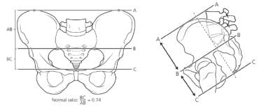
    Calculation of the sacral ratio. Calculation of the sacral ratio.
    Spinal ultrasonography in the newborn period and up to age 3 months (at which point the sacrum ossifies) may be performed to find evidence of a tethered spinal cord and other spinal anomalies.
    Cross-table lateral radiography may help demonstrate the air column in the distal rectum in the small percentage of patients in whom clinical evidence does not delineate the likely anorectal anomaly in 16-24 hours.

    Imaging Studies After Newborn Period

    High-pressure distal colostography is performed on an outpatient basis, after the colostomy has been created. Hydrosoluble contrast material is injected into the distal stoma to demonstrate the precise location of the distal rectum and its likely urinary communication.
    Hydrostatic pressure under fluoroscopic control is required. A Foley catheter is placed in the mucous fistula, and the 3-cm3 balloon is inflated and pulled back to occlude the stoma during contrast injection. The hydrostatic pressure must be high enough (manual syringe injection) to overcome the muscle tone of the striated muscle mechanism that surrounds the rectum and keeps it collapsed. This is the best way to demonstrate a rectourinary communication and determine the rectum's true height.
    The contrast material usually fills the proximal urethra and bladder through the fistula. The injection is continued until the child voids, and pictures are taken during micturition to reveal, in a single picture, the sacrum, rectum height, perineum, fistula location, bladder, vesicoureteral reflux (if present), and urethra. When colostography is performed correctly, voiding cystography and cystoscopy are not necessary.
    Colostography is vital in determining the anatomy to plan definitive repair. In 10% of patients, the fistula is at the level of the bladder neck; in these instances, during the main repair, the surgeon knows that the rectum can be found only through the abdomen, and a combined posterior sagittal and abdominal or laparoscopic approach is used.
    The anorectal defect of imperforate anus without fistula may also be demonstrated with this radiologic evaluation. Occurring in approximately 5% of patients, imperforate anus without fistula has a good functional prognosis and is common in individuals with Down syndrome.
    In most newborn girls with anorectal malformations (except for those with persistent cloaca), distal colostography is not necessary because the fistula is clinically evident.
    If the spine was not evaluated with ultrasonography in the newborn period, magnetic resonance imaging is necessary after age 3 months to exclude the presence of tethered cord and other spinal anomalies.
  • Approach Considerations

    Indications for surgical treatment (colostomy vs anoplasty) are discussed more fully elsewhere (see Presentation). No absolute contraindications to surgery exist. The presence of associated malformations, such as prematurity and congenital cardiac disease, often postpones definitive colorectal reconstruction.
    Potential methods of evaluation for anorectal malformations, including prenatal diagnosis and genetic karyotyping to reveal familial disposition, are areas of rapid advance.

    Medical Therapy

    Associated malformations must be addressed.

    Genitourinary defects

    Approximately 50% of all patients with anorectal malformations have an associated urogenital anomaly, which commonly varies with the type of anorectal defect.
    A list of anorectal defects and the percentages of patients with associated urogenital anomalies is as follows:
    • Persistent cloaca - 90%
    • Rectobladderneck fistula - 84%
    • Rectoprostatic urethral fistula - 63%
    • Rectovestibular fistula - 47%
    • Rectobulbar urethral fistula - 46%
    • Rectoperineal fistula - 26%
    • Imperforate anus without fistula - 31%
    All patients must be examined at birth for these defects; the most valuable screening test is abdominal and pelvic ultrasonography. Urologic evaluation prior to colostomy provides the surgeon the necessary information to address the urologic problem if needed.

    Tethered cord

    A tethered spinal cord refers to the intravertebral fixation of the phylum terminale. Tethered cord has a known association with anorectal malformation; approximately 25% of patients with anorectal malformation have a tethered spinal cord. The prevalence of this anomaly increases with increasing height and complexity of the anorectal anomaly. In addition, patients with a hypodeveloped sacrum and associated urologic problems are more likely to have tethered cord. Motor and sensory disturbances of the lower extremities may result.
    Patients with anorectal malformations and tethered cord have a poorer prognosis for bowel and urinary function; they also have higher anorectal defects, less-developed sacra, other spinal problems, and less-developed perineal musculature. The actual impact of tethered cord alone on functional prognosis remains unclear.
    The neurosurgical literature indicates that untethering the cord avoids motor and sensory problems. No evidence suggests that this operation affects the functional prognosis of patients with anorectal malformation, but may improve bladder emptying capacity.
    Spinal ultrasonography in the first 3 months of life and magnetic resonance imaging (MRI) thereafter are useful radiologic modalities to establish the diagnosis.

    Sacral and spinal defects

    The sacrum is the most commonly affected bony structure. Traditionally, the number of sacral vertebral bodies was counted to evaluate the degree of sacral deficiency. A more objective assessment of the sacrum may be obtained by calculating a sacral ratio. The sacrum is measured and its length is compared with bony parameters of the pelvis (see the image below). The lateral view is more accurate than the anteroposterior view because its calculation is not affected by pelvic tilt.
    Calculation of the sacral ratio. Calculation of the sacral ratio.
    Assessment of sacral hypodevelopment correlates with the patient's functional prognosis. Normal sacra have a calculated sacral ratio greater than 0.7. Bowel control has rarely been observed in patients with calculated sacral ratios of less than 0.3. Hemisacrum is almost always associated with a presacral mass (teratomas or anterior meningoceles). Hemivertebrae may also affect the lumbar and thoracic spine, predisposing to scoliosis.
    Patients may have spinal anomalies other than tethered cord, such as syringomyelia and myelomeningocele.

    Surgical Therapy

    As discussed previously, the surgeon must decide in the newborn period whether the child requires fecal diversion with a colostomy or if a primary procedure is possible.

    Colostomy

    A descending colostomy with separated stomas is recommended. The advantages of this type of colostomy include the following:
    • Only a small portion of distal colon is defunctionalized, but with an adequate amount of rectosigmoid for the future pullthrough
    • Washing and cleaning the portion of the colon distal to the colostomy is relatively easy
    • Distal colostography is easy to perform
    • The separated stomas prevent spillage of stool from proximal to distal bowel, which avoids impacted distal stool and urinary tract infections
    • Prolapse with this technique is uncommon; proximal stoma prolapse in a normally rotated colon should not occur with this technique, because the colon is well fixed to the retroperitoneum just before the colostomy rises to skin level; because the distal stoma is in a mobile portion of the colon, it may prolapse; the distal stoma must intentionally be made small, both to avoid prolapse and because it is used only for irrigations and radiologic studies
    When performing a colostomy in the newborn, the distal bowel should be irrigated to remove all the meconium. This prevents formation of a megasigmoid, which may lead to constipation after the colostomy ultimately gets closed.
    Colostomy errors include the following:
    • Too-distal sigmoidostomy - In this most common error, the colostomy is placed too distal and interferes with the pullthrough procedure
    • Right upper sigmoidostomy - Instances of inadvertent sigmoid colostomy placed in the right upper quadrant during an attempt to perform a transverse colostomy have occurred. Inadvertent anchoring of the sigmoid in the right upper quadrant interferes with the pullthrough procedure
    • Incomplete diversion of stool - A loop colostomy does not divert the stool completely and allows for distal stool impaction and urinary tract infections
    • Megarectum - Transverse colostomies may produce megarectum, due to passage and accumulation of mucus

    Definitive repair

    Repair of an anorectal malformation requires a meticulous, delicate technique and a surgeon with experience in treating these defects. The posterior sagittal approach is ideal for defining and repairing anorectal anomalies.
    Anorectal abnormalities in 90% of newborn boys may be repaired solely with a posterior sagittal approach, whereas 10% require an additional abdominal component (with laparotomy or laparoscopy) to mobilize a very high rectum. All anorectal malformations in newborn girls may be repaired with the posterior sagittal approach, with the exception of approximately 30% of instances of persistent cloaca. In this 30%, the rectum or vagina is high enough to also require an abdominal approach.
    A Foley catheter is placed first; then, patients are placed in the prone position with the pelvis elevated.
    The posterior sagittal incision length varies depending on the anorectal defect. Perineal fistulas are repaired with a minimal posterior sagittal incision that is large enough to divide the external sphincter and to mobilize the anus back to the center of the sphincter complex. The sphincter mechanism is always located posterior to the fistula site. This operation may be performed in the neonatal period without a protective colostomy.
    The posterior sagittal approach is based on the fact that nerves do not cross the midline. Remaining exactly in the midline, the surgeon preserves the innervation of all the important pelvic structures.
    An electrical stimulator helps reveal the location of the sphincteric mechanism. The parasagittal fibers, the muscle complex, and the levators are identified during the dissection. The external sphincter is represented by muscle fibers that run parallel to the midline in a parasagittal fashion. A muscle structure termed the levator mechanism lies medial to these fibers and represents the lower end of the funnel-like voluntary muscle.
    The levator mechanism extends in continuum down to the skin, but this was not known prior to the use of the posterior sagittal approach. Electrically stimulated, the parasagittal fibers elicit a contraction that results in shortening of the same fibers. The rectum pushes forward toward the pubic bone when the upper portion of the levators are stimulated. Stimulating the lower part of the funnel-like muscle structure elevates the anal dimple. This group of muscle fibers, termed the muscle complex, extends from the levator mechanism down to the skin and is located immediately medial to the parasagittal fibers.
    The surgeon opens posterior sagittally. The skin and subcutaneous tissue are divided, and the parasagittal fibers below are divided in the midline, as is the muscle complex. The levator muscle is then opened, and the rectum is found, except in patients with a true supralevator malformation (10% of cases), in whom the surgeon will instead find a genitourinary structure with this approach.
    When the rectum is located, its posterior wall is opened in the midline to demonstrate the presence of a fistula. This posterior incision in the rectum is carried down to the fistula site. The rectum and urethra share a common wall. Meticulous dissection is required to separate the distal rectum from the urethra. A submucosal dissection must be performed in the first 5 mm above the fistula site.
    The rectum is separated from the urinary tract. This dissection is facilitated through placing multiple 6-0 silk sutures in the rectal mucosa to exert uniform traction. The opening in the urethra is then closed with absorbable suture.
    Once the rectum is separated, it is then mobilized down to reach the perineum by circumferentially dividing the bands and vessels that hold the rectum up in the pelvis. The intramural blood supply of the rectum is excellent; therefore, the rectum remains viable. Because the newborn depends on this intramural blood supply, preventing damage to the rectal wall is vital.
    Once the rectum is fully mobilized, the size of the rectum and the available space must be assessed. The rectum occasionally requires tapering to fit the limits of the sphincteric mechanism. The tapering should take place on the posterior wall so that the suture lines of the tapered rectum and the closed urethral fistula do not lie next to each other.
    The rectum is placed in the limits of the sphincter mechanism, which is reconstructed around it in the midline. The rectum is sutured to the perineal skin (anoplasty).
    In 10% of newborn boys with this defect, the rectum enters the urinary tract at the bladderneck level. The repair of this malformation involves a posterior sagittal incision and an abdominal component. The distal rectum is separated from the urinary tract, mobilized, and pulled through to lay within the sphincteric funnel. The pathway is just under the coccyx and sacrum in the area of the pelvic retroperitoneum.
    To mobilize the rectum off of the bladderneck, an abdominal component is required via laparoscopy or laparotomy. The fistula is ligated with great care to avoid injuring the ureters and vas deferens: the plane of dissection must be made close to the bowel wall of the rectosigmoid, 2-3 cm above the peritoneal reflection. The rectum is then mobilized for adequate length to reach the perineum without tension.
    The fistula is divided and sutured with absorbable material. The rectum is passed through to the posterior sagittal incision and an anoplasty is performed. This part can be done with legs lifted up, remaining in supine position.
    In patients with imperforate anus without fistula, the same meticulous dissection is required to separate the distal rectum from the urinary tract as in patients with rectourinary fistulae, because the rectum and urethra still share a common wall.
    In patients with rectovestibular fistula, the posterior sagittal incision may be shorter than in newborn boys with rectourethral fistulae. Often, the entire levator mechanism does not need to be divided; only the external sphincter, muscle complex, and part of the lower portion of the levator mechanism require division. The rectum and posterior vagina share a common wall; this separation is the most difficult part of the operation. Once the rectum is completely mobilized, the perineal body is constructed, and the rectum is placed within the limits of the sphincter mechanism.
    A rare malformation, rectal atresia, occurs in 1% of patients. The anal canal is normal, and, externally, the anus appears typical. However, a blockage exists 1-2 cm from the anal skin and is usually found when the nurse tries to pass a thermometer. These babies should undergo colostomy at birth; definitive repair involves a posterior sagittal approach and an anastomosis between the posterior rectum and the anal canal. This defect is particularly associated with a presacral mass, which must be sought.

    Postoperative Details

    Postoperative management

    The posterior sagittal incision is relatively painless.
    In patients with a rectourethral fistula, the Foley catheter usually stays in place for approximately 5-7 days.
    If the colostomy is untouched during the operation and laparotomy or laparoscopy was not necessary, oral feedings may be started immediately postoperatively. If a laparotomy or laparoscopy was necessary, the patient may require a period of fasting and nasogastric decompression.
    At 2 weeks after surgery, anal calibration is performed, followed by a program of anal dilatations. The anus must be dilated twice daily, and the  size of the dilator is increased every week. The final size to be reached depends on the patient’s age. Once the desired size is reached, the colostomy may be closed. Dilatations are continued afterward according to a prescribed protocol. Dilatations are a vital part of postoperative treatment to avoid an anoplasty stricture.
    After colostomy closure, severe diaper rash is common, because the perineal skin has never before been exposed to stool.

    Postoperative functional disorders

    Constipation is the most common problem encountered after treatment for imperforate anus. It is the most important problem to avoid after definitive repair in newborn girls with rectovestibular or rectoperineal fistula and in newborn boys with rectobulbar urethral fistula, imperforate anus without fistula, and rectoperineal fistula. Failure to avoid constipation may result in megarectum and megasigmoid and can lead to fecal impaction and overflow incontinence. SeeBowel Management for further discussion.
    The origin of the constipation problem is unknown. Originally, the perirectal dissection was believed to cause a degree of denervation that resulted in constipation. However, on careful review of the largest patient series, those with the most benign defects (ie, the least amount of perirectal dissection) experienced the worst constipation.
    The presence of a megarectum prior to the pull-through procedure correlates with postoperative constipation. Megarectum is more common in patients who underwent a transverse or loop colostomy during the newborn period.
    Constipation appears to be a hypomotility disorder secondary to chronic bowel dilatation; alternatively, the hypomotility may cause the dilatation. Dilatation causes constipation, creating a vicious cycle. Patients who have undergone an older operation, an abdominoperineal operation for imperforate anus that included rectum resection, are prone to develop diarrhea because of lack of a rectal reservoir. Incontinence in these patients is much more difficult to treat because stool constantly passes (see Bowel Management).
    Occasionally, constipation becomes so severe that patients develop chronic fecal impaction and constant soiling. These patients are often referred to as having fecal incontinence. However, if the patient has a type of anorectal anomaly with a good prognosis, this incontinence is often overflow pseudoincontinence. Once the constipation is treated, the patient regains continence.
    When constipation is severe and the patient has a megasigmoid (and the patient is fecally continent), resection of the sigmoid has been found to dramatically reduce the patient's laxative requirements. The descending colon with normal caliber and motility is anastomosed to the rectum at the peritoneal reflection. This procedure is useful for the select group of patients who require enormous amounts of daily laxatives to keep their colons clean. The rectal reservoir should be preserved to avoid the problem of diarrhea-related incontinence.

    Complications

    Complications of surgery include dehiscence and infection, which may be avoided with colostomy before the main repair. These complications may compromise the chance of achieving typical bowel function.
    With inadequate preoperative anatomic information, the urinary tract is at considerable risk because the surgeon does not know the precise anorectal defect and if the surgeon approaches a low rectum transabdominally, they risk leaving behind the very distal rectum, which becomes a posterior urethral diverticulum.

    Long-Term Monitoring

    The key in these patients is to treat constipation proactively and, if possible, avoid it after the pull-through procedure altogether. Patients must be regularly monitored, and laxatives and dietary manipulations are begun at the first sign of constipation.
    Patients may experience soiling. This may represent fecal incontinence in patients with very high imperforate anus or in those with poor muscles and an abnormal sacrum. These patients require a bowel management program (see Bowel Management). However, in a patient with a good prognosis, soiling may represent overflow incontinence, and constipation must be treated.
    The child’s bowel movement pattern before toilet-training may provide important information concerning the potential for continence. For example, a 1-year-old child who has undergone a pullthrough procedure for imperforate anus and has one to three discrete bowel movements per day has good potential for future fecal continence. Signs of feeling are demonstrated while the child is pushing during a bowel movement.
    On the other end of the spectrum, a child who has fecal incontinence passes stool constantly without evidence of pushing or feeling. Whereas a child with a typical bowel movement pattern is trainable, a child with the pattern of fecal incontinence likely requires a bowel management program with a daily enema. The child with true fecal incontinence should not be expected to achieve voluntary bowel control.